Datoteka:Pterosaur respiratory system.jpg
Stranica ne postoji na drugim jezicima.
Izvor: Wikipedija

Vidi sliku u punoj veličini(3.006 × 4.057 piksela, veličina datoteke: 2,52 MB, MIME tip: image/jpeg)


Logo Wikimedijinog spremnika Ova je datoteka sa Zajedničkog poslužitelja i mogu je rabiti drugi projekti. Opis s njezine stranice s opisom datoteke prikazan je ispod.
Opis

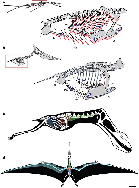
Models of ventilatory kinematics and the pulmonary air sac system of pterosaurs.

a, Model of ventilatory kinematics in Rhamphorhynchus. Thoracic movement induced by the ventral intercostal musculature results in forward and outward displacement of the distal vertebral and proximal sternal ribs, and ventral displacement of the sternum, upon inspiration (blue arrows and pink outline). In addition, ventral expansion of the abdomen is induced through caudoventral rotation of the prepubis. Ranges of skeletal movement were modelled after those observed in vivo in the avian thorax and the crocodylian pelvis [26], [27]. Rhamphorhynchus modified from Wellnhofer [48]. b, Model of ventilatory kinematics in Pteranodon wherein the fused anterior vertebral ribs and articulation of the scapulocoracoid with the supraneural plate and anterior sternum limit movement of the anterior sternum, which cannot undergo elliptical rotation. However, the posterior vertebral ribs, sternal ribs, sternum, and prepubis are still capable of anterodorsal-posteroventral excursions facilitating volumetric increases and decreases of the thorax during inspiration-expiration. Pteranodon modified from Bennett [29]. c, d, reconstruction of pulmonary air sac system in the Lower Cretaceous ornithocheirid Anhanguera santanae (AMNH 22555). c, Lateral view showing the inferred position of the lungs (orange), cervical (green) and abdominal air sacs (blue), as predicted on the basis of postcranial skeletal pneumaticity. Thoracic air sacs (shown in grey) are also likely to have been present, but generally do not leave a distinct osteological trace. Humerus and more distal forelimb not shown. d, Dorsal view illustrating the inferred position of subcutaneous diverticular networks (light blue) distally along the wing. The right side depicts a conservative estimate for the size of the airsac network, limiting it to the pre-axial margin of the wing based solely on the presence of pneumatic foramina in closely positioned wing bones. The left side depicts the likely maximal size of an inferred diverticular network, accounting for its inclusion between the dorsal and ventral layers of the wing membrane. Scale = 10 cm. Skeletal reconstruction in c, d modified from Wellnhofer [49]. Abbreviations: as in figure 2, and: Cor: coracoid portion of scapulocoracoid, Ga: gastralia.
Datum
Izvor http://www.plosone.org/article/info%3Adoi%2F10.1371%2Fjournal.pone.0004497;jsessionid=A57F0FDB595AC49992E2B5A390FA104C
Autor Leon P. A. M. Claessens, Patrick M. O'Connor, David M. Unwin
w:hr:Creative Commons
imenovanje autora
Ova datoteka je licencirana pod licencijom Creative Commons Imenovanje 2.5 nelokalizirana licencija.
Slobodno smijete:
  • dijeliti – umnožavati, distribuirati i javnosti priopćavati djelo
  • remiksirati – prerađivati djelo
Pod sljedećim uvjetima:
  • imenovanje autora – Morate pripisati odgovarajuće autorske zasluge, dati poveznicu na licenciju, te naznačiti jesu li načinjene promjene autorskog djela. Prethodno navedeno možete učiniti na svaki razuman način, ali ne na način koji bi sugerirao da Vi ili Vaše korištenje licencorova djela ima izravno licencorovo odobrenje.
PLOS
PLOS
This file was published in a Public Library of Science journal. Their website states that the content of all PLOS journals is published under the Creative Commons Attribution 4.0 license (or its previous version depending on the publication date), unless indicated otherwise.

Opisi

Dodajte kratko objašnjenje što predstavlja ova datoteka

Predmeti prikazani u ovoj datoteci

motiv

Povijest datoteke

Kliknite na datum/vrijeme kako biste vidjeli datoteku kakva je tada bila.

Datum/VrijemeMinijaturaDimenzijeSuradnikKomentar
sadašnja23:17, 2. ožujka 2009.Minijatura za inačicu od 23:17, 2. ožujka 2009.3.006 × 4.057 (2,52 MB)FunkMonk{{Information |Description=Models of ventilatory kinematics and the pulmonary air sac system of pterosaurs. a, Model of ventilatory kinematics in Rhamphorhynchus. Thoracic movement induced by the ventral intercostal musculature results in forward and out

Nijedna stranica ne rabi ovu datoteku.

Globalna uporaba datoteke

Sljedeći wikiji rabe ovu datoteku:

Metapodatci