
Datoteka:Nematocyst discharge.png
Viša rezolucija nije dostupna.
Nematocyst_discharge.png (480 × 371 piksela, veličina datoteke: 190 KB, MIME tip: image/png)
| Ova je datoteka sa Zajedničkog poslužitelja i mogu je rabiti drugi projekti. Opis s njezine stranice s opisom datoteke prikazan je ispod. |

|
This biology image could be re-created using vector graphics as an SVG file. This has several advantages; see Commons:Media for cleanup for more information. If an SVG form of this image is available, please upload it and afterwards replace this template with
{{vector version available|new image name}}.
It is recommended to name the SVG file “Nematocyst discharge.svg”—then the template Vector version available (or Vva) does not need the new image name parameter. |
Sažetak
| Opis |
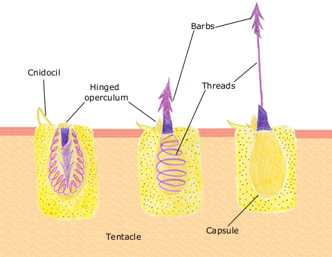
English: The diagram above shows the anatomy of a nematocyst cell and its “firing” sequence, from left to right. On the far left is a nematocyst inside its cellular capsule. The cell’s thread is coiled under pressure and wrapped around a stinging barb. When potential prey makes contact with the tentacles of a polyp, the nematocyst cell is stimulated. This causes a flap of tissue covering the nematocyst—the operculum—to fly open. The middle image shows the open operculum, the rapidly uncoiling thread and the emerging barb. On the far right is the fully extended cell. The barbs at the end of the nematocyst are designed to stick into the polyp’s victim and inject a poisonous liquid. When subdued, the polyp’s tentacles move the prey toward its mouth and the nematocysts recoil back into their capsules. |
| Datum | 11. travnja 2007. (izvorni datum postavljanja) |
| Izvor | Prebačeno s en.wikipedia na Zajednički poslužitelj . |
| Autor | Izvorno postavio Spaully na Wikipediji na engleskom jeziku |
Licencija
This file is licensed under Creative Commons ShareAlike 1.0 License.
Creative Commons has retired this legal tool and does not recommend that it be applied to works.
|
Ova slika je u javnom vlasništvu jer sadrži materijale koji su izvorno iz Nacionalne oceanske i atmosferske administracije SAD, koja je nastala fotografiranjem ili izradom kao dio službenih dužnosti zaposlenika.
|
Izvorna evidencija postavljanja
Izvorna stranica s opisom bila je ovdje. Niže navedena suradnička imena odnose se na en.wikipedia.
- 2007-04-11 17:10 Spaully 480×371×8 (194868 bytes) Modified from: http://www.oceanservice.noaa.gov/education/kits/corals/media/supp_coral01b.html {{Information |Description=Nematocyst discharge process. |Source= Modified from [http://www.oceanservice.noaa.gov/education/kits/corals/media/supp_coral01b.html
Povijest datoteke
Kliknite na datum/vrijeme kako biste vidjeli datoteku kakva je tada bila.
| Datum/Vrijeme | Minijatura | Dimenzije | Suradnik | Komentar | |
|---|---|---|---|---|---|
| sadašnja | 19:29, 13. listopada 2007. | | 480 × 371 (190 KB) | Alison | {{Information |Description===Description== The diagram above shows the anatomy of a nematocyst cell and its “firing” sequence, from left to right. On the far left is a nematocyst inside its cellular capsule. The cell’s thread is coiled under pressur |
Uporaba datoteke
Na ovu sliku vode poveznice sa sljedećih stranica:
Globalna uporaba datoteke
Sljedeći wikiji rabe ovu datoteku:
- Uporaba na ca.wikipedia.org
- Uporaba na ceb.wikipedia.org
- Uporaba na en.wikipedia.org
- Uporaba na fr.wikipedia.org
- Uporaba na id.wikipedia.org
- Uporaba na it.wikibooks.org
- Uporaba na ja.wikipedia.org
- Uporaba na lv.wikipedia.org
- Uporaba na ms.wikipedia.org
- Uporaba na my.wikipedia.org
- Uporaba na pa.wikipedia.org
- Uporaba na pt.wikipedia.org
- Uporaba na simple.wikipedia.org
- Uporaba na sv.wikipedia.org
- Uporaba na te.wikipedia.org
- Uporaba na th.wikipedia.org
- Uporaba na vi.wikipedia.org